


Surgical Dermatology

Skin Biopsies
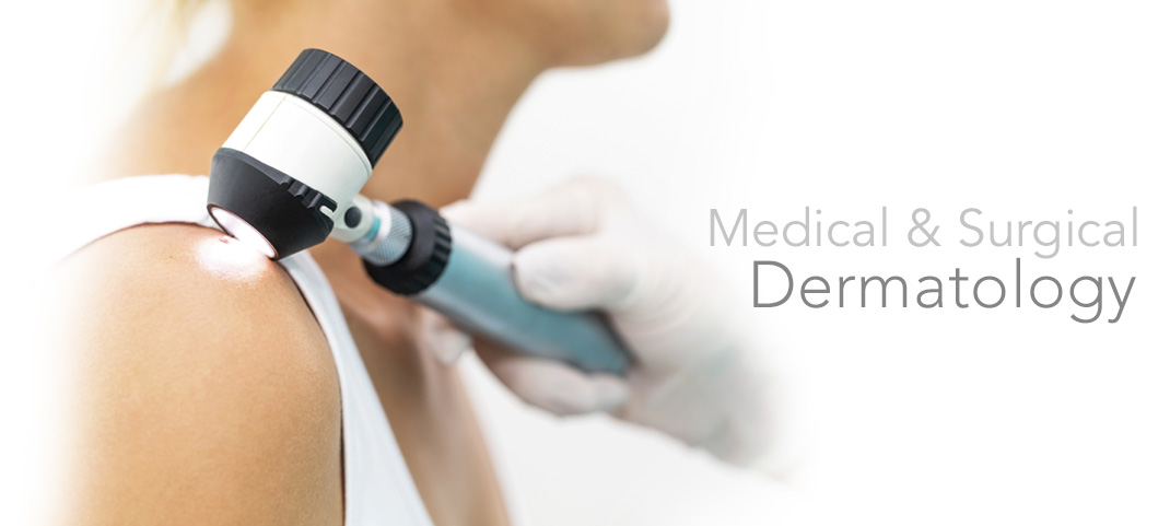
A skin lesion biopsy is a diagnostic test that involves removing a tissue sample and examining it under a microscope. This test is used to identify suspicious lesions and to differentiate normal cells from abnormal ones.
Before undergoing a skin biopsy, the physician should be notified if the patient has a bleeding disorder, is taking any blood-thinning medication, or has an autoimmune disorder or underlying skin condition. Since a local anesthetic will be administered prior to the biopsy, the procedure will not be painful.
Following the biopsy, the tissue sample is stored in a preservative solution or a sterile container until it can be processed at a pathology laboratory. Once the specimen has been prepared and thoroughly examined under a microscope, a diagnosis will made to determine what the course of follow-up treatment, if any, should be.
Removal of Moles
Many moles and birthmarks on the skin are completely benign, and do not pose a health threat. Oftentimes, though, people want these moles removed because they find them unattractive. Types of moles include dysplastic nevi, which can become melanomas. Dysplastic nevi are usually irregular in size, shape, color and border. They can be located on any area of the body, not only those exposed to the sun.
Depending on their depth, location and color, as well as the patient's skin type, age and other factors, treatment for benign but unattractive moles and birthmarks may use the following methods.
Laser or Pulsed-Light Therapy: Laser or pulsed-light therapy is most beneficial on superficial moles and birthmarks. Laser mole removal is only able to treat flat superficial moles. Moles with deep roots cannot be removed effectively with a laser, and may grow back.
Surgical Excision: Surgical excision may be considered for deep or raised moles or birthmarks. It may also be recommended for irregular or potentially malignant moles, which will include a biopsy of mole tissue. During an excision, the doctor cuts out the entire mole and surrounding tissue, and stitches the skin closed.
Surgical Shave: Surgical shaving is recommended for smaller moles and does not require stitches. The doctor numbs the area around the mole and uses a surgical blade to cut around and beneath the mole.
Removal of Skin Cancers
Skin cancer is the most common form of cancer in the United States. It involves abnormal growths of skin cells that can form anywhere on the body, but most frequently appear in areas that are regularly exposed to the sun such as the skin of the face, head and neck. In addition to excessive sun exposure, certain factors, such as fair skin, moles and aging, can also increase the risk of skin cancer.
Mohs micrographic surgery is a safe and effective treatment for skin cancer that thoroughly excises the tumor while only mildly disturbing surrounding tissue. It is the only skin cancer treatment available that targets just cancerous tissue through comprehensive microscopic examination of the affected area.
Mohs surgery excises not only the visible tumor but also any roots that may have extended beneath the surface of the skin. Five-year cure rates have been demonstrated up to 99 percent for first-treatment cancers and 95 percent for recurring cancers.
This procedure is most commonly used for the treatment of basal and squamous cell carcinomas, although it can also be used to treat melanoma and other types of cancer. Mohs surgery is often recommended for recurring cancer because its results are so thorough. It is also ideal for treating cancer in cosmetically and functionally prominent areas such as the face, head and neck.
Destruction of Actinic Keratoses
An actinic keratosis, also known as a solar keratosis, is a common premalignant skin lesion. An actinic keratosis occurs when the cells that comprise 90 percent of the epidermis, the keratinocytes, change their size, shape or organization in a process called cutaneous dysplasia.This alters the texture of the skin surface and may extend deeper, into the dermis.
Depending on the location and severity of the lesion, an actinic keratosis may be treated in a number of ways. The patient and doctor will decide on methodology in consultation. These may include:
- Cryotherapy, or freezing
- Curettage, or scraping
- Application of cream or ointment
- Chemical peeling
- Photodynaminc therapy using laser light
Acne Surgery (Unclog Pores)
Acne is a common condition that causes pimples, cysts and other lesions to develop on the skin of the face, neck, chest, back, shoulders and upper arms. Acne is caused by clogged sebaceous glands, which can lead to blocked pores. Acne develops on the skin when the pores become clogged, as a result of an overproduction of oil. When oil builds up in the hair follicle it forms a soft plug that forces the follicle wall to bulge and protrude from the skin, causing a lesion to develop.
If cystic acne is painful and bothersome to patients, a dermatologist may perform a procedure called "drainage and extraction" to remove a large acne cyst. This procedure may also be performed when the cyst has not responded to oral or topical medication. Draining and extracting the cyst also helps to relieve pain and reduces the chance that the cyst will leave a scar.
The dermatologist performs the extraction using a special metal instrument that presses down firmly on the targeted area of the skin to gently unclog the plug and remove any oil, bacteria or debris from the lesion. For certain types of blemishes, a sterile needle may be used to break the skin before removing any debris or buildup from the area. Extractions may also be performed with steaming techniques, which help to open the pores of the skin and minimizes the trauma to the skin. After the extraction procedure, some patients may experience temporary redness or swelling which can be relieved by applying cold compresses to the skin.
Removal of Cyst
Cysts are balloon-like structures in the skin filled with solid or fluid material. They most often contain sebaceous material, the oily substance that would normally be present on the surface of the skin for normal lubrication. Cysts can occur anywhere on the body, although the face, neck, back and area behind the ears are the most common sites. They develop as an infection, often from a swollen hair follicle, and require treatment to prevent them from enlarging or becoming cancerous.
Cyst removal is typically done through surgical excision. A small incision is made in the area of the cyst and then the cyst and surrounding tissue will be removed to ensure complete excision. A local anesthetic is used for this procedure. Most cysts do not return when thoroughly removed. Some patients are left with a small scar after a cyst is removed, which can be further treated with a reconstructive treatment, although most scars will fade over time.
Cyst Drainage
Cyst aspiration removes fluid from a fluid-filled nodule called a cyst. During the procedure, the skin is sterilized and a local anesthetic is administered. Once the area is numb, a needle is inserted into the patient's skin, often using ultrasound guidance. The tip of the needle is placed in the cyst and fluid is withdrawn until the cyst is emptied and collapses. It is important that the wall of the cyst be completely removed in order to avoid regrowth.
No bandages are required after cyst aspiration, and there should be no aftereffects. Cyst aspiration takes less than 5 minutes to perform. The fluid extracted from a cyst is sent to a pathology lab to confirm that the cyst is benign.
Keloid & Scar Revision
Keloids are overgrown areas of scar tissue that form at the site of a previous injury such as an incision, wound, vaccination, pimple or piercing. They appear on the skin as an irregularly-shaped pink or red scar that is raised above the rest of the skin and continues to grow into areas that were not affected by the initial injury.
Treatment of keloids depends on the personal preference and desired outcome of the patient. The only true cure for keloids is to prevent them from occurring, but there are several procedures available to help improve the appearance and related symptoms. Some of these treatments help flatten keloids, while others reduce redness and size. Most treatments will leave an irregular mark or texture on the skin.
Keloid treatments include:
- Cortisone injections
- Cryotherapy
- Interferon
- Laser removal
- Surgery
Although keloids are not medically dangerous, many people seek treatment to restore the appearance of their skin and help the keloid become less noticeable over time. Your doctor can help you decide which treatment option is best for you.
Surgical Removal of Moles
Not all Moles are created equal. Requires physical examination and biopsy, if necessary